We investigate gross morphology and microscopic anatomy of animals using image analysis, histology and immunohistochemistry, isotropic fractionator for cell quantification, geometric morphometrics.
Olfactory system of fish
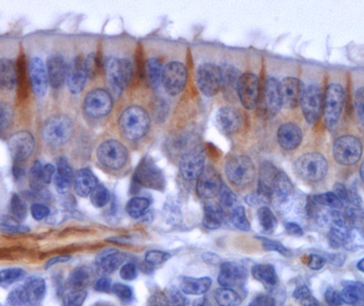
Chemoreception is the most ancient sense. The olfactory system of vertebrates has several intriguing features: the sensory epithelium hosts the olfactory neurons, which are continuously renewed during the whole lifespan of the animal, and that are directly in contact with the environment. These neurons project their axons, that form the olfactory nerve or cranial nerve I, to a region of the brain called olfactory bulb. They form structures that are unique to the bulb: the glomeruli. There are no glomeruli in the rest of brain and, in such a peculiar structure, the olfactory stimuli are at first elaborated.
The polyphyletic group of fish presents a great wondrous variety in habitats, behaviors, life strategies, and morphological adaptations. Fish olfactory system is not an exception and its comparative study is a fascinating window on chemoreception evolution.

Fish brain in numbers
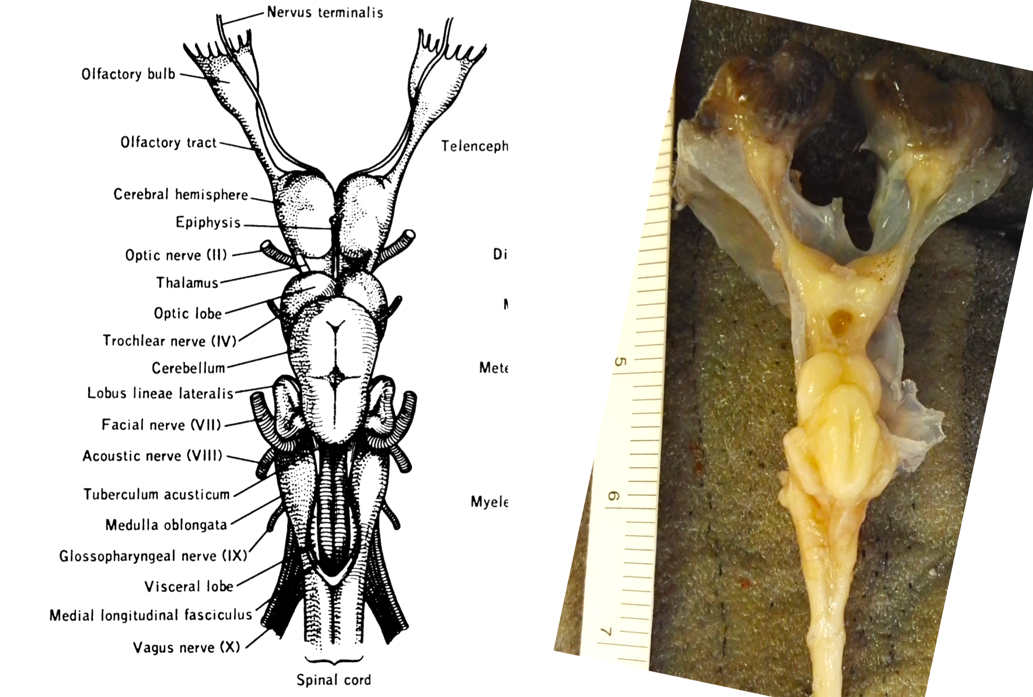
The anatomy of the central nervous system can be studied at different levels of detail. The comparison of relative or absolute mass or volume of brain regions, from different species, has been a great deal of interest in the study of evolution and ecomorphology. Techniques that allow to quantify the number of neurons and glial cells brought this kind of study to a further level. At the Coalab we apply these techniques to fish brain.

Elasmoid scales morphology
Scales are remarkable structures on the fish body. Their shape and size are extremely variable among species and along the fish body. We apply geometric morphometrics, both landmarks-based and outlines-based, to analyze shape variation in teleost elasmoid scales.
